Introduction to Sample Preparation and Library QC for Ion Torrent™ Sequencing
Publication Date: September 2015 (tpub_164)
Abstract
In this interview, we spoke with Dr. Molly Accola of the University of Wisconsin. Dr. Accola holds a Masters in Medical Science from Harvard Medical School and a Ph.D. in Immunology from Harvard University, with a post-doctoral residency at Mayo Clinic. She worked as an R&D scientist at Third Wave Technologies, Inc. (now Hologic), and Pharmaceutical Product Development, LLC (PPD), both in Madison, WI. She has been the Senior Laboratory Development Specialist at the University of Wisconsin Hospital and Clinics since 2007, during which she has helped develop many assays. She has most recently started using next-generation sequencing to research potentially pathogenic variants in solid tumors.
Introduction
Q: Which NGS system do you use? When you were evaluating various systems, how did you decide on the platform you chose?
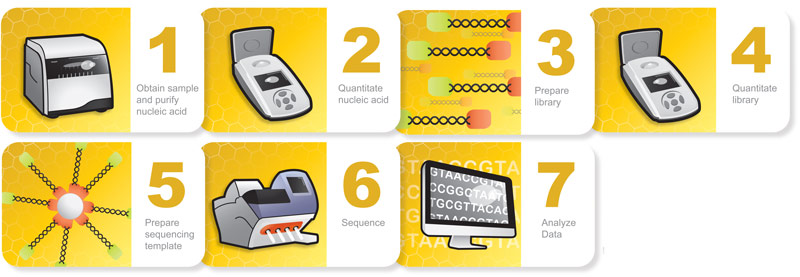
A: That’s a simple answer—it’s cost. Essentially we looked at the Illumina MiSeq and we looked at the Ion Torrent PGM™. The PGM and the OneTouch™ systems together were less than $100,000, which fit our budget. The workflow is also kit-based (Figure 1). Life Tech has a panel of oligos that’s prepackaged to target 50 oncology-associated genes. Their protocol, although a little tedious, is very straight forward and easy to use for a lab just starting NGS, so that’s why we chose the PGM.
Q: Could you describe the general process used in your lab of preparing and conducting NGS?
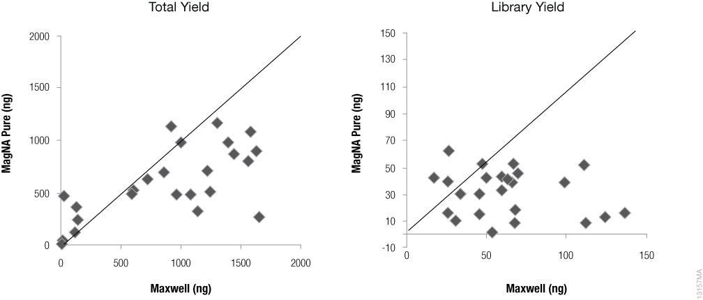
A: When we want to sequence a sample, we obtain three FFPE tissue slides, one of which (the middle one) is H&E (Hematoxylin and Eosin) stained. Macro-dissection is the first step. We put an H&E slide up next to an unstained slide and circle the area that we want to extract DNA from. We scrape that area off of the unstained slides and put that into the tube with mineral oil, which is the beginning of the Maxwell® CSC extraction. What we found was that as long as there was sufficient tumor and tissue, we could get away with using only one slide for the Maxwell® CSC for DNA extraction (Figure 2).
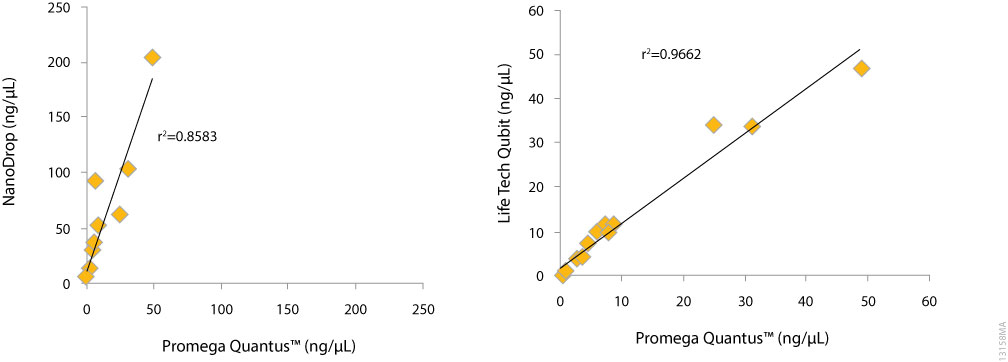
Once extracted, we have to quantify the sample for NGS. For sample quantitation we use the Quantus™ Fluorometer, which is more accurate at the lower end (than absorbance), and that’s invariably where we are with FFPE. The Life Tech fluorometer is the Qubit™, though we find that the Quantus™ Fluorometer works equally well (Figure 3). The cost is also a little bit less per sample with the QuantiFluor® dye. Specifically we have been using the QuantiFluor® ONE dsDNA System. We like that because the pre-diluted format takes one step away from the assay setup, so now it’s one fewer step than running the Qubit™ assay. Ultimately, you need to have a very good quantitation estimate before you start the library prep—you’re targeting a normalization input of 10ng. This is where less is probably better than more, because if you find that your library prep is unsuccessful the last thing you want to do is use more DNA in the reaction. So, quantification before library preparation is critical.
It usually takes a full day to macro dissect, extract on the Maxwell® instrument, and quantify before we are ready to do library preparation the following day. We like to get all of the library prep, including a whole series of different incubations in the thermocycler, done in one day so that by the end of the day we can quantify the library. And I should mention too, after library preparation, performing an additional library quantification is key. This is important because the ratio of DNA to Ion Sphere™ Particles (or beads) for the template preparation will dictate whether the emulsion PCR is clonal (and can be sequenced on the PGM) or polyclonal. Adding too much DNA to the emulsion PCR increases the polyclonal reads, which are not interpretable by the instrument and are discarded. Adding too little DNA will reduce the number of reads obtained for the sample because there will be more 'bald' or untemplated beads (Ion Sphere™ Particles); there is unused real estate on the chip. Sometimes we start the template preparation that same day, which I think is great. I don’t want to push people or throw too much at someone when they are starting next generation sequencing, because it’s a lot just to figure out how to use your time most efficiently, but it really is very smooth to get to that library quantification and start the template prep which will go over night on the Ion One Touch™ instrument.
The beginning of the third day we will enrich the library, initialize the PGM, load the chip and we’re off and running. So if we start working on a sample Monday morning we can have it ready by Wednesday. And actually, if that’s the first chip we run we can get results Wednesday afternoon.
Q: What bioinformatics software packages do you use?
A: We use IGV (Integrated Genomics Viewer), and if the particular variation does not have a COSMIC (Catalog of Somatic Mutations in Cancer) number, we then determine if it is a coding change. We also use Cartagenia software which really helps annotation of variants, especially those that don’t have COSMIC numbers. This allows us to decipher whether it’s a variant of unknown significance, whether it’s pathogenic, whether it’s benign and so on.
Q: How has your NGS workflow or success rate changed as a result of using fluorescent quantitation methods such as the Quantus™ fluorometer and QuantiFluor® ONE system?
A: Accurate nucleic acid quantification is essential to successful NGS. Instruments using UV spectroscopy, although common in the laboratory, are not sufficiently accurate. In our hands, quantifying by UV spec gives results that average greater than five times the concentration given by fluorescence. This is likely to have a huge impact on library prep and downstream NGS because you wouldn't be putting enough DNA into the reactions, resulting in significantly fewer reads. Fluorescent quantification methods, such as the Quantus™ and QuantiFluor® ONE, not only achieve the required accuracy for NGS, but they are fast, easy, and relatively inexpensive, too. These are important characteristics considering that the NGS workflow is quite time consuming and costly.