How to Measure Target Degradation
Quantitative measurement of target protein levels fuels degrader discovery and optimization while deepening our understanding of cell biology and disease. We offer these complementary approaches:
- HiBiT technology allows ultrasensitive detection of protein degradation for high-throughput screens; HiBiT knock-in cell lines work seamlessly with both endpoint and live-cell kinetic assays to monitor degrader compound properties
- Lumit® Immunoassay Cellular Systems use a labeled-antibody approach to quantify target-protein levels, allowing degrader efficacy measurements across a broad range of relevant cell backgrounds

How Do You Measure Protein Degradation with Knock-In Cell Lines?
Measure Degrader Activity with HiBiT Knock-In Cell Lines
HiBiT is an 11-amino-acid peptide tag, which has high affinity for its complementary partner, LgBiT. When bound together, they form a binary luminescent protein that produces a bright, sensitive signal proportional to target protein levels. You can quantify HiBiT-tagged proteins with luminescence or the Anti-HiBiT Monoclonal Antibody.
Measuring degrader activity using HiBiT is simple:
- Introduce HiBiT at the endogenous locus using CRISPR.
- Add degrader compound to trigger protein degradation, causing luminescence signal to drop.
- Measure the signal loss in real time or at the endpoint to calculate precise degrader potency and kinetics.

The HiBiT protein tagging system accommodates lytic, extracellular and intracellular live-cell detection.
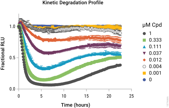
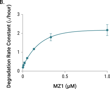
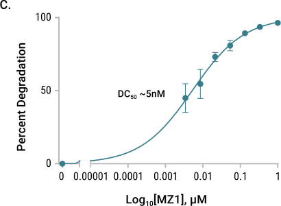
Degradation kinetics of endogenous HiBiT-BRD4 following PROTAC treatment. HiBiT was inserted at the endogenous BRD4 locus in the HEK293 LgBiT Cell Line. Cells were treated with a titration of MZ1 in CO2-independent medium containing Nano-Glo® Endurazine™ Substrate. Panel A. Kinetic luminescence; Panel B. Degradation rate; C: Dmax.
Download this white paper to see more data: Advancing Quantitative Analysis of Targeted Protein Degrader Compounds.
How to Measure HiBiT Protein Degradation: Endpoint and Live-Cell Analysis
To start, generate a stable cell line expressing HiBiT-tagged target protein using CRISPR/Cas9 knock-in. This enables you to study the target protein under endogenous expression conditions.
Endpoint Analysis
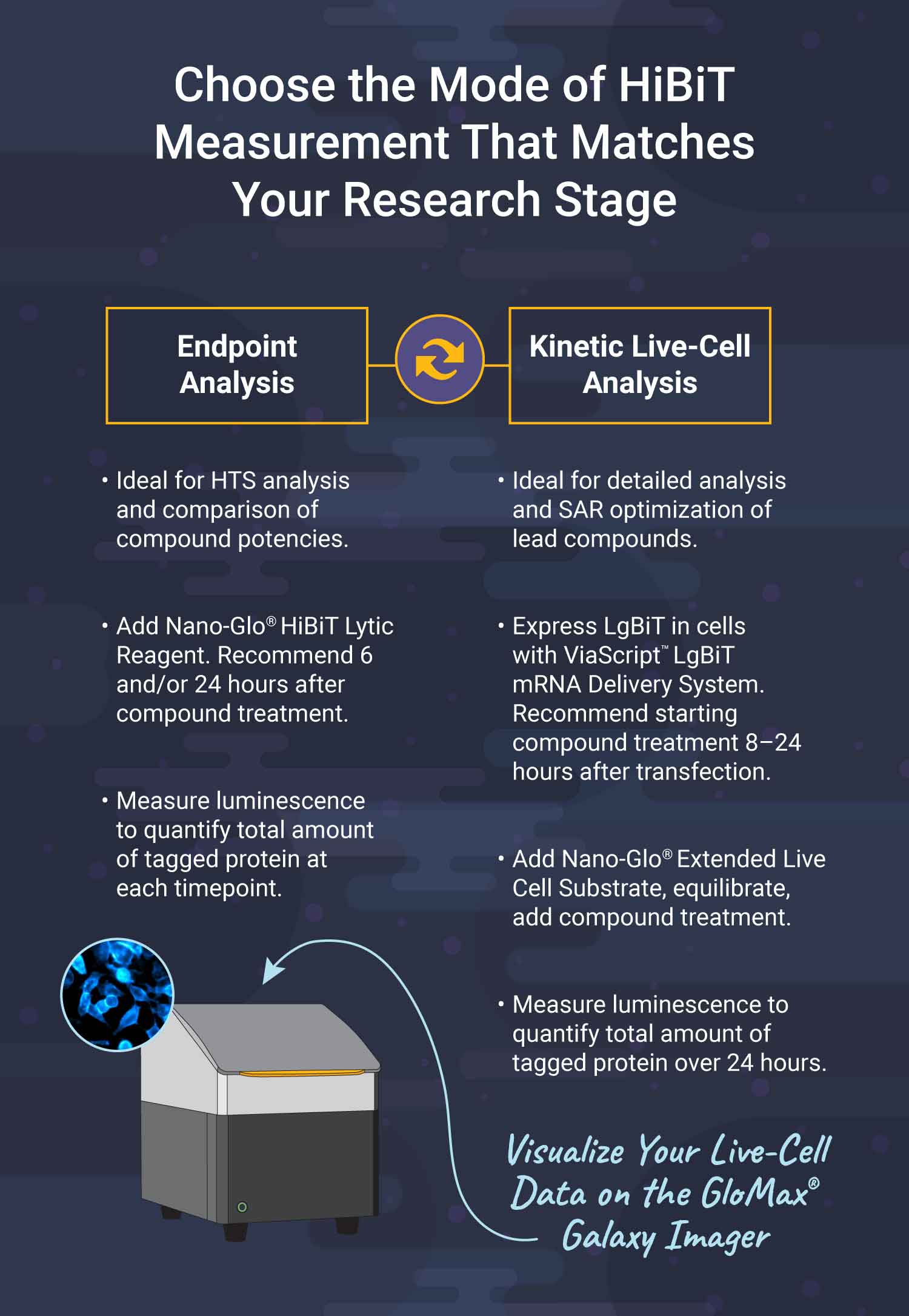
Measure HiBiT-tagged protein levels at fixed time points to quantify degrader compound efficacy with high-throughput. Generate cellular dose–response curves to calculate DC50/Dmax values and assess both on-target efficacy and off-target degradation.
Kinetic Live-Cell Analysis
Monitor HiBiT-tagged protein degradation in real time:
- Express LgBiT intracellularly using ViaScript® LgBiT mRNA Delivery System for rapid uniform expression across cell lines.
- Detect luminescence in real time using Nano-Glo® Extended Live Cell Substrates on the GloMax® Discover instrument.
- Analyze kinetic data to derive compound parameters, such as degradation rate and duration at Dmax. Analyze results faster using the Pronect™ TPD Kinetic App.
How Do You Measure Protein Degradation in Native Cells?
Monitoring Targeted Protein Degradation with Lumit® Immunoassays
Lumit® Immunoassays utilize split-luciferase technology, in which analyte sensors (like antibodies, antigens, or binding proteins) are chemically labeled with two luciferase subunits. These two subunits only produce a luminescent signal when they come together in close proximity, which happens when the antibodies bind to their target analyte.
Why Use Lumit® Immunoassays to Detect Protein Degradation?
- Speed & Simplicity: Simple add-mix-read protocol with no wash steps
- Native Cell Models: Quantifies protein degradation in disease-relevant cell backgrounds
- High-Throughput Capability: Enables rapid screening of multiple degrader compounds and cell lines simultaneously
- Sensitive & Quantitative: Detects both direct protein degradation and downstream signaling changes with high sensitivity
- Flexibility: Works with any protein target provided two robust antibodies are available.
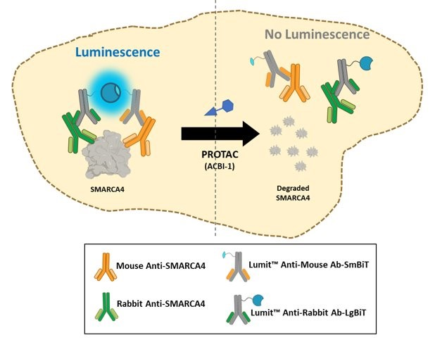
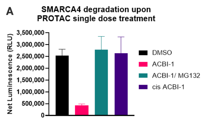
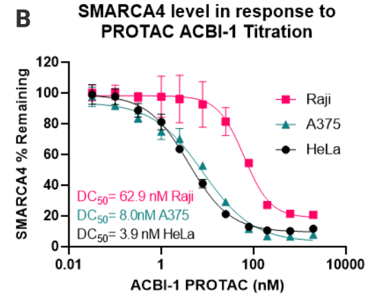
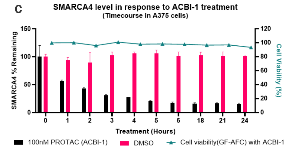
Degradation of native SMARCA4 following PROTAC treatment. 50,000 cells were seeded per well. After treatment with the indicated compounds, the Lumit® Immunoassay Cellular System (Set 2) was used to detect the level of SMARCA4. Panel A. SMARCA4 level after treatment with 250nM of ACBI-1, 250nM ACBI-1and 20µM MG132, 250nM Cis-ACBI-1, or DMSO for 5 hours. Panel B. Profiling SMARCA4 degradation with ACBI-1 in three different cell lines. Panel C. Time course of SMARCA4 degradation with ACBI-1.
Need help with Endpoint or Kinetic Degradation Profiling?
Learn more about our targeted protein degradation services.